DNA barcoding technology is able to easily mark an unprecedented number of connections between individual brain cells. The unexpected complexity of the visual system is only the first of its secrets.
Sitting at the desk in his office on the campus of
Cold Spring Harbor , the neuroscientist
Tony Zador turned his computer monitor to me to show a complex graph in the form of a matrix. Imagine a spreadsheet that is filled with colors of various shades and gradations instead of numbers. In passing, he said: “When I tell people that I understood the connections of tens of thousands of neurons and show them that, in response, they just say“ A? ”But when I show people this ...” He pressed a button and a transparent screen appeared three-dimensional model of the brain, rotating around one of the axes, filled with nodes and lines, in an amount too large to be counted. "They say:" What the ...! "
Zador showed me a map of 50,000 neurons in the cortex of the mouse. It marked where the bodies of all neurons are located, and where they direct their long axon branches. Neural maps of this size and detail have never been before. Zador abandoned the traditional approach of mapping the brain through fluorescent neuron markup, and chose an unusual technology based on the long tradition of molecular biology research at Cold Spring Harbor on Long Island. He used particles of genome information to insert a unique RNA sequence, or barcode, into each individual neuron. Then he cut the brain into cubes and fed them to a DNA sequencer. The result was a three-dimensional image of 50,000 neurons of the mouse brain cortex (and soon more will be added to them), with resolutions up to single cells.
This work,
magnum opus Zadora, is still undergoing a series of refinements and revisions before publication. But in a recent paper published in Nature magazine, they and their colleagues demonstrated that this technique, known as MAPseq (Multiplexed Analysis of Projections by Sequencing), can be used to search for new cell types and patterns previously unknown . It was also shown that this high-performance markup method is a serious competitor in the accuracy of the fluorescent technique, which is the current standard, but works best with only a small number of neurons.
 Tony Zador
This project was born thanks to Zadora’s dissatisfaction with his routine “core” work as a neurophysiologist, how he speaks dryly of her. He studies the effect of hearing on decision-making in rodents: how their brain hears sounds, processes audio information and determines a behavioral response or action. Electrophysiological recordings and other traditional instruments used to solve such questions did not satisfy the scientist who is prone to mathematics. The problem, as Zador says, is that neural connections are not well known to us, therefore, as a part-time job, he is trying to create new tools for imaging the brain.
The current state of advanced brain-mapping technology is the
Allen Brain Atlas brainstorming project, assembled as a result of several years of work by many laboratories, and it cost about $ 25 million. The Allenov Atlas is known as the mass communication atlas, as it tracks group connections. It is very useful to researchers, but it is not able to make subtle differences that exist within groups or subpopulations of neurons.
If we ever want to know how a mouse hears a high trill, processes it and understands that sound means a reward in the form of a refreshing drink, or forms a new memory, to later remember the threat, we need to start with a map or brain connections . From Zdor’s point of view, a lack of knowledge about such compounds plays a role in why progress in the treatment of psychiatric diseases is so slow, and why artificial intelligence is still not intelligent enough.
Justus Kebschul , a neuroscientist at Stanford University, the author of a new work for Nature and a former graduate student at Zadora’s laboratory, said that doing neuroscience without knowing about the connections is like “trying to understand how a computer works, looking at it from the outside, stuffing his electrode and trying to understand what can be found there. Not knowing that the hard disk is connected to the processor, and USB is transmitting input to the system, it is very difficult to understand what is happening. "
Inspiration for the development of SAPsek came to Zador, when he learned about another brain mapping technology called
Brainbow [coloring of nerve cells with fluorescent proteins in different colors; brain - brain, rainbow - rainbow / approx. trans.]. This method, which appeared in the laboratory of
Jeff Lichtman of Harvard University, is notable for being able to genetically mark up to 200 individual neurons at the same time using different combinations of fluorescent dyes. The result was a picturesque multi-color
picture of neuronal neurons, showing in detail a complex mixture of axons and bodies of neurons. This revolutionary work gave hope that the markup of the
connectome — a complete description of all the nervous connections of the brain — was approaching reality. Unfortunately, the limitation of this technology in practice was that, when viewed through a microscope, experimenters could recognize from five to ten different colors, which was not enough to penetrate into the intricacies of neurons in the cerebral cortex and markup of many neurons at the same time.
It was then that an idea arose in Zadora’s head. He realized that the problem of the extraordinary complexity of a connectom can be tamed if the researchers succeed in adapting the increasing speeds and diminishing costs of high-yield genome sequencing technologies for their needs. “In mathematics, this is called reducing the problem to the previous one, which has already been solved,” he explained.
Researchers introduce genetically modified viruses into the animal that carry many known RNA sequences or “barcodes”. For the order of the week, viruses multiply in the animal's body, and fill each neuron with a unique combination of these bar codes. When researchers then cut the brain into pieces, RNA barcodes can help them track individual neurons from piece to piece.
Zador's idea led to the emergence of a
new work in Nature, in which his laboratory and a team from University College in London, led by neurobiologist
Thomas Mersik-Flegel, used SAPsek to track the connections of nearly 600 neurons in the mouse's visual system.
600 neurons - a modest start compared to the tens of millions of neurons contained in the mouse brain. But this was enough for a specific goal that the researchers set themselves. They wanted to see if there was a structure in the brain connections pattern that could shed light on his work. A popular theory is that in the visual cortex, individual neurons collect individual pieces of information from the eyes - about the edges of objects in sight, or the type of movement, or spatial orientation, for example. The neuron then sends a signal to one appropriate area of the brain that specializes in processing this type of information.
An example of how SAPsec can determine the connections of multiple neurons:
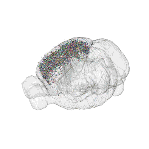 Colored dots indicate the location of the bodies of 50,000 neurons on the cortex of the mouse
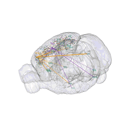 Axonal connections of only two neurons, ending somewhere else in the brain
 Nerve pathways of multiple neurons
To test his theory, the team first tagged several mouse neurons in the traditional way, injecting genetically encoded fluorescent dye into individual cells. Then, they used a microscope to track how the cells stretched from the main visual cortex to other parts of the brain. They found that the axons of the neurons branched and sent information to several brain regions at once, which refuted the one-to-one connection theory.
Then they began to look for patterns of these connections. They used SAPsec to track the connections of 591 neurons that branched and innervated various targets. The team saw that the distribution of axons is subject to regularities: some neurons always extend axons into areas, say, A, B and C, and never into areas D or E.
The results show that the visual system has extremely complex connections and that the patterns of these connections are much more complicated than just a one-to-one relationship. “High-level visual areas do not just receive the information specially processed for them,” Kebskul said. Instead, many sites receive the same information, “so their calculations can be related to each other.”
However, the fact that certain cells have connections to certain areas also means that there are specialized, not yet open cells in the visual cortex. Kebskul said that this map is similar to the blueprints that will allow future researchers to understand what these cells are doing. “SAPsek allows you to build an equipment map. As soon as we deal with the equipment, we will be able to start working with the software and the calculation process, ”he said.
The competitive advantage of SAPsec in terms of speed and cost is very significant. According to Zador, this technology should be able to increase its scale to 100,000 neurons, processing such volume in a couple of weeks and for only $ 10,000 - this is much faster than traditional methods of markup and much cheaper than them.
Such advantages will make the task of marking and comparing the nerve paths of many brains a more realistic task. The study of conditions such as
schizophrenia and
autism , which are believed to arise from differences in brain connection patterns, often frustrated scientists, because the tools available to them are not able to recognize many small details about neural connections. One can imagine that researchers will be able to build a map of these states for mice and compare them with more typical examples of brain maps that will spur a new wave of research. "Many mental illnesses are caused by problems at the level of connections," said
Hon Kui Zen , executive director of the structured sciences department at the Allen Institute for the Study of the Brain. "Relationship information will tell you where to look for answers."
Detailed markup will also allow scientists to collect a large amount of neurobiological data and look for patterns among them that reflect the general principles of the brain. “Tony stares at the brain with an undistorted look,” said
Shrikant Chalasani , a molecular neuroscientist at the Salk Institute. “As the human genome map provided support for testing hypotheses and searching patterns in sequences and genes, the Tony method can do the same for brain architecture issues.
A detailed map of the human genome did not instantly explain all the mysteries of the work of biology, but it gave a list of biomolecular "spare parts" and opened the way for a
whole pleiad of revolutionary research . Similarly, at the current stage of development, SAPsec cannot provide information about the work or location of the cells it marks, or show which cells communicate with each other. But Zador is planning to add such functionality soon. It also works in conjunction with scientists studying various parts of the brain, such as neural connections that handle fear.
“I think that many ideas will be drawn from all these connections. But this, as with the genome - in itself it is not something interesting, the things that it allows to do are revolutionary. Therefore I am in such joyful excitement, - told Zador. “I hope that this technology will provide support for the next generation of work in this area.”